Publicaciones

Identifying injury: “A retrospective cohort study based on information and communication technology”.

Publicado en: Archives of Orthopaedic and Trauma Surgery

Referencias:

Maryem I.; Bejarano, P; Villafañe, J.H, Ph.D, Msc; Lopez-Alcorocho, J.M.; Fernandez-Jaén, T.F., Guillén-Vicente, I.; Guillén-Garcia, P. “Identifying

injury: “A retrospective cohort study based on information and communication technology”. Archives of Orthopaedic and Trauma Surgery

----------------------

Case Report: Efficacy of dobesilate in insertional Achilles tendinopathy

Publicado en: F1000research

Leer más:

Achilles tendinopathy is an overuse syndrome, common among runners, with sometimes considerable negative impact on their performance, overall health, and well-being. Our report shows that local injection of an aqueous solution of the diethylammonium salt of dobesilate, an inhibitor of fibroblast growth factor with significant anti-angiogenic and anti-inflammatory effects, is effective in reducing vascular density and pain in insertional Achilles tendinopathy.


Referencia:

Cuevas P., Fernández-Jaen, T., Guillén, P. Angulo, J.y Giménez-Gallego, G. “Case Report:Efficacy of dobesilate in insertional Achilles tendinopathy” F1000research (2016), doi: 10.12688/f1000research.7260.1


Link: https://f1000research.com/articles/5-34/v1

----------------------

Viability of pathologic cartilage fragments as a source for autologous chondrocyte

Publicado en: Cartilage


OBJECTIVE:

To study if a culture of chondrocytes can be obtained from pathologic hyaline cartilage (PHC) fragments.

DESIGN:

Twenty-five men and 9 women with osteochondritis dissecans (OCD) in 11 cases, arthrosis in 13 patients, and trauma in the remaining 10 cases were included. The PHC fragments and a small sample of the next healthy cartilage were extracted by arthroscopy. According to the appearance, the PHC samples were divided into fixed (3 cases), flapped (6 patients), or loose bodies (25 cases), depending on the attachment degree of the cartilage to the subchondral bone. Approximately half of each pathologic sample and the whole healthy one were digested to isolate the cells trying to establish the cell culture.

RESULTS:

We were able to establish a cell culture in 7 out of 34 (20.6%) PHC samples (positive samples), whereas in the remaining 27 (79.4%) no cell growth was observed (negative samples). Most of the negative samples were loose bodies (P = 0.005) taken from patients with OCD or arthrosis (P = 0.001) with an evolution time of more than 1 year (P < 0.001). The best binary logistic regression model (P < 0.001) showed that the only factor affecting the establishment of cell culture was the evolution time (P = 0.044).

CONCLUSION:

It is possible to culture chondrocytes from osteochondral fragments if they are traumatic, within a year of injury and not from fragments due to arthrosis or OCD.


Referencia:

Guillen García, P., Rodriguez Iñigo, E. Guillén Vicente, I, Guillén Vicente, M., Fernández Jaén, T. Concejero López, V. Val, Maestro, A. Abelow, S. y López-Alcorocho, J.M. “Viability of pathologic cartilage fragments as a source for autologous chondrocyte” Cartilage  Vol. 7(2) 149-156 DOI: 10.1177/1947603515621998. SAGE (2016).


Link: https://www.ncbi.nlm.nih.gov/pmc/articles/PMC4797241/


----------------------

Nuestra experiencia con la técnica de implante de condrocitos autólogos para el tratamiento de lesiones condrales: resultados de 50 pacientes a 2 años de seguimiento

Publicado en: Rev. Esp. Artroscopia y Cirugía Articular.


Aim

The aim of this work was to evaluate the clinical results of the matrix-induced autologous chondrocyte implantation (MACI) technique on chondral defects of the knee and ankle. A study was conducted on 150 patients treated with MACI (performed by the same team of surgeons in all cases) during 2002-2007.

Methods

The lesion was evaluated by arthroscopy, and a cartilage biopsy was taken. The cellular implant was performed by arthroscopy in 53 patients, and by open surgery in the remaining cases. The results were measured following an in-house clinical protocol. Data corresponding to a mean follow-up period of 24 months were available in 50 out of the 150 implanted patients. Histological studies were performed in a second biopsy taken by arthroscopy in 5 patients 2 years after cell implantation.

Results

In 89% of patients without previous surgeries, and in 66% of patients with previous surgeries, the results were good or excellent for pain and motion, and most of them were able to return to their previous physical activities, including professional sport. Histological studies performed on a second biopsy showed that the new tissue is hyaline-like cartilage with lower cell numbers and subjectively softer than normal cartilage.

Conclusion

Arthroscopic MACI is a promising procedure to treat chondral lesions in the knee and ankle, although the histological analysis suggests that increasing cellular dosage would improve the results of the technique.

Clinical relevance

The importance of this study is that it presents an increased number of patients with chondral lesion treated with MACI. A high number of these patients were treated with arthroscopic MACI. In the present study it is shown that the treatment gives good results.


Referencia:

Guillen Garcia, P., Rodriguez Iñigo, E., Arauz de Robles, S., Guillén Vicente, M. Guillen Vicente, I. Caballero Santos, R., Ramos Martin, T., Fernández Jaén, T. Abelow, S. y J.M. López-Alcorocho. “Nuestra experiencia con la técnica de implante de condrocitos autólogos para el tratamiento de lesiones condrales: resultados de 50 pacientes a 2 años de seguimiento”. Rev. Esp. Artroscopia y Cirugía Articular.  22(3):120-125 (2015)


Link: http://www.sciencedirect.com/science/article/pii/S2386312915000833

----------------------

Spanish Consensus Statement: The Treatment of Muscle Tears in Sport

Publicado en: Orthopaedic Journal of Sports Medicine


On the 21st of March, 2015, experts met at Clínica CEMTRO in Madrid, Spain, under the patronage of The Spanish Society for Sports Traumatology (SETRADE), The Spanish Federation of Sports Medicine (FEMEDE), The Spanish Association of Medical Services for Football Clubs (AEMEF), and The Spanish Association of Medical Services for Basketball Clubs (AEMB) with the aim of establishing a round table that would allow specialists to consider the most appropriate current general actions to be taken when treating muscle tears in sport, based on proven scientific data described in the medical literature. Each expert received a questionnaire prior to the aforementioned meeting comprising a set of questions concerning therapeutic indications generally applied in the different stages present during muscle repair. The present Consensus Document is the result of the answers to the questionnaire and resulting discussion and consensus over which are the best current indications in the treatment of muscle tears in sport. Avoiding immobilization, not taking nonsteroidal anti-inflammatory drugs (NSAIDs) randomly, fostering early mobilization, increasing vascularization of injured, site and regulating inflammatory mechanisms-without inhibiting these from the early stages of the recovery period-all stood out as main points of the Consensus Document. Additionally, there is controversy concerning cell stimulation techniques and the use of growth factors or cell inhibitors. The decision concerning discharge was unanimous, as was the criteria considered when it came to performing sport techniques without pain.


Referencia:

  1. Fernández Jaén et all. “Spanish Consensus Statement: The Treatment of Muscle Tears in Sport” Published December 30, 2 2015 doi: 10.1177/2325967115622434.Orthopaedic Journal of Sports. Medicine Decembre Vol. 3, 12 (2015)


Link: https://www.ncbi.nlm.nih.gov/pmc/articles/PMC4710119/

----------------------

Infrared thermography as a support tool for screening and early diagnosis in emergencies

Publicado en: Journal of Medical Imaging and Health Informatics


Sufficient methods to reliably diagnose sport injuries and to specify the lesions produced exist; however, these methods are not simultaneously real-time, low-cost, harmless and easy-to-use as screening methods for emergency trauma services. This study attempts to establish the capacity of infrared thermography (IRT) to discriminate injuries and to evaluate its applicability in emergency trauma scenarios. The sample consisted of 201 patients in the Emergency Unit at the CEMTRO clinic in Madrid (108 males and 93 females). The average and maximal temperatures of the skin (T sk) from the injured and uninjured region of interest (ROI) were collected from a thermogram registered with a T335 FLIR infrared camera, and the results were compared with the diagnosis of the emergency unit practitioner. The T sk differences between the injured and the uninjured areas (T sk) were significant (p < 0.05) for both the average (ΔT sk = +0.5 °C) and maximum (T sk = +0.6 °C) temperatures. These side-to-side ΔT sk data agree with the thresholds of asymmetry found in previous studies. The results of this pilot study, arranged by injured ROI, type of injury, medical diagnosis of the practitioner and evolution time of the injury, showed that IRT had a good specificity for detecting temperature asymmetries on injured areas. The influence of the usage of ice and anti-inflammatory creams on the ΔT sk results was analysed using some of the excluded cases from the general study. The results of this article indicate that thermography is a valid tool to determinate the existence of an injury. When a high resolution infrared imager is used and an appropriate protocol is followed, IRT can be a good support tool, providing practitioners with additional information to correctly identify a sport injury.


Referencia:

  1. Sillero Quintana, T. Fernández Jaén, I. Fernandez Cuevas, P.. Gómez Carmona, J. Arnaiz Lastras, M.D. Pérez Pérez y P. Guillén García. “Infrared thermography as a support tool for screening and early diagnosis in emergencies” American Scientific Publishers. Journal of Medical Imaging and Health Informatics. Vol. 5, 1-6 (2015).


Link: http://www.ingentaconnect.com/content/asp/jmihi/2015/00000005/00000006/art00013

----------------------

The importance of the intercondylar nothc in anterior cruciate ligament tears

Publicado en: The Orthopaedic Journal of Sports Medicine

BACKGROUND:

The factors associated with anterior cruciate ligament (ACL) tears are not completely clear. Some studies have shown that patients with a narrow intercondylar notch have a predisposition for ACL tears.

PURPOSE:

To determine the relationship between the α angle and intercondylar notch width measurements and ACL tears.

STUDY DESIGN:

Case-control study; Level of evidence, 3.

METHODS:

A total of 530 patients (308 with ACL rupture, 222 with healthy ACLs) were included in this study. The α angle and intercondylar width were measured from magnetic resonance images (MRIs). Binary logistic regression analysis was performed to determine the influence of the variables on ACL status (normal or torn). Odds ratios (ORs) and their respective 95% CIs were also calculated.

RESULTS:

No significant differences in patient age and the affected knee were found between patients with normal or torn ACLs. The mean α angle was higher in patients with a torn ACL than in those with an intact one (57.5° ± 5.5° vs 56.2° ± 4.5°; P = .009). Intercondylar width was significantly lower in patients with a torn ACL than in those with an intact one (18.2 ± 3.1 vs 19.5 ± 3.6 mm; P < .001). A highly significant difference between men and women was found for mean intercondylar notch width (19.3 ± 3.3 vs 17.4 ± 3.1 mm; P < .001). In a logistic regression model, sex, intercondylar width, and α angle were statistically significant when adjusted for age.

CONCLUSION:

Study results suggest that the ACL tears are associated with a narrow intercondylar notch and a high α angle, and that tears occur more frequently in men than in women.

CLINICAL RELEVANCE:

The model proposed in this study could be used by the physician in the medical office as a tool to identify the risk factors that may predispose a patient for a potential ACL tear.


Referencia:

  1. Fernández-Jaén, J.M. Lopez-Alcorocho, E. Rodriguez-Iñigo, F. Castellan, J.C. Hernández y P. Guillen-García. “The importance of the intercondylar nothc in anterior cruciate ligament tears” The Orthopaedic Journal of Sports Medicine, 3(8), 2325967115597882 DOI: 10.1177/2325967115597882 (2015).


Link: https://www.ncbi.nlm.nih.gov/pmc/articles/PMC4622305/


----------------------

Overouse injuries in profesional ballet, injury-based Differences among ballet disciplines

Publicado en: The Orthopaedic Journal of Sports Medicine

BACKGROUND:

Despite overuse injuries being previously described as the most frequent in ballet, there are no studies on professional dancers providing the specific clinical diagnoses or type of injury based on the discipline.

HYPOTHESIS:

Overuse injuries are the most frequent injuries in ballet, with differences in the type and frequency of injuries based on discipline.

STUDY DESIGN:

Cross-sectional study; Level of evidence, 3.

METHODS:

This was a descriptive cross-sectional study performed between January 1, 2005, and October 10, 2010, on injuries occurring in professional dancers from leading Spanish dance companies who practiced disciplines such as classical, neoclassical, contemporary, and Spanish ballet. Data, including type of injury, were obtained from specialized medical services at the Trauma Service, Fremap, Madrid, Spain.
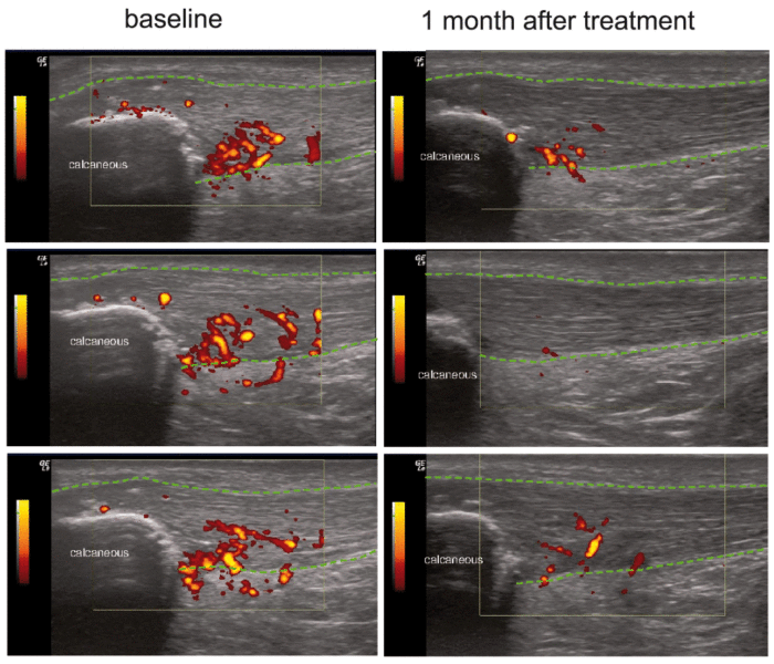
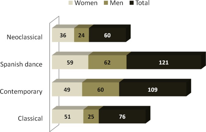
RESULTS:

A total of 486 injuries were evaluated, a significant number of which were overuse disorders (P < .0001), especially in the most technically demanding discipline of classical ballet (82.60%). Injuries were more frequent among female dancers (75.90%) and classical ballet (83.60%). A statistically significant prevalence of patellofemoral pain syndrome was found in the classical discipline (P = .007). Injuries of the adductor muscles of the thigh (P = .001) and of the low back facet (P = .02) in the Spanish ballet discipline and lateral snapping hip (P = .02) in classical and Spanish ballet disciplines were significant.

CONCLUSION:

Overuse injuries were the most frequent injuries among the professional dancers included in this study. The prevalence of injuries was greater for the most technically demanding discipline (classical ballet) as well as for women. Patellofemoral pain syndrome was the most prevalent overuse injury, followed by Achilles tendinopathy, patellar tendinopathy, and mechanical low back pain.

CLINICAL RELEVANCE:

Specific clinical diagnoses and injury-based differences between the disciplines are a key factor in ballet.


Referencia:

F.J. Sobrino, C. de la Cuadra y P. Guillén, “Overouse injuries in profesional ballet, injury-based Differences among ballet disciplines”. The Orthopaedic Journal of Sports Medicine, 3 (6), 2325967115590114. DOI: 10.1177/2325967115590114  (2015).

Link: https://www.ncbi.nlm.nih.gov/pmc/articles/PMC4622371

---------------------- ----------------------