Archives for 12 Abr,2017

You are browsing the site archives by date.

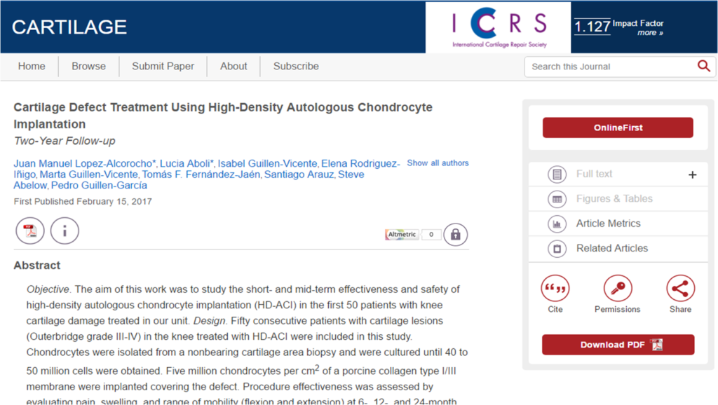
Cartilage Defect Treatment Using High-Density Autologous Chondrocyte Implantation: Two-Year Follow-up

Publicado en: Cartilage

Objective. The aim of this work was to study the short- and mid-term effectiveness and safety of high-density autologous chondrocyte implantation (HD-ACI) in the first 50 patients with knee cartilage damage treated in our unit. Design. Fifty consecutive patients with cartilage lesions (Outerbridge grade III-IV) in the knee treated with HD-ACI were included in this study. Chondrocytes were isolated from a nonbearing cartilage area biopsy and were cultured until 40 to 50 million cells were obtained. Five million chondrocytes per cm2 of a porcine collagen type I/III membrane were implanted covering the defect. Procedure effectiveness was assessed by evaluating pain, swelling, and range of mobility (flexion and extension) at 6-, 12-, and 24-month follow-up. The International Knee Documentation Committee (IKDC) subjective evaluation form was used to evaluate symptoms and functions of the knee. Results. The percentage of patients with pain and swelling decreased progressively in the following visits, with differences being statistically significant (P < 0.001 and P = 0.040, respectively). IKDC scores improved progressively throughout the 24-month follow-up (P < 0.001). Thus, the mean IKDC score improvement was 26.3 points (95% confidence interval [CI] = 18.2-34.4 points) at 12 months and 31.0 points (95% CI = 22.9-39 points) at 24 months. No significant differences were found when performing extension (P = 0.112). Flexion significantly improved by 25.1° at 24-month follow-up (P = 0.013). Conclusions. HD-ACI is a safe and effective technique for the treatment of cartilage defects, improving clinical and subjective perception of knee functionality. These preliminary results encourage future studies comparing this technique with traditional ACI.

Referencia:

J.M. López-Alcorocho, L. Aboli, I. Guillén-Vicente, E. Rodriguez Iñigo, M. Guillén-Vicente, T. F. Fdez.-Jaén, S. Arauz, S. Abelow and P. Guillén-García. “Cartilage Defect Treatment Using High-Density Autologous Chondrocyte Implantation: Two-Year Follow-up” Cartilage 1-7 DOI:10.1177/1947603517693045


Link: http://journals.sagepub.com/doi/abs/10.1177/1947603517693045


----------------------

Interspecies chimerism with mammalian pluripotent stem cells

Publicado en: Cell


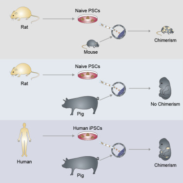
Interspecies blastocyst complementation enables organ-specific enrichment of xenogenic pluripotent stem cell (PSC) derivatives. Here, we establish a versatile blastocyst complementation platform based on CRISPR-Cas9-mediated zygote genome editing and show enrichment of rat PSC-derivatives in several tissues of gene-edited organogenesis-disabled mice. Besides gaining insights into species evolution, embryogenesis, and human disease, interspecies blastocyst complementation might allow human organ generation in animals whose organ size, anatomy, and physiology are closer to humans. To date, however, whether human PSCs (hPSCs) can contribute to chimera formation in non-rodent species remains unknown. We systematically evaluate the chimeric competency of several types of hPSCs using a more diversified clade of mammals, the ungulates. We find that naïve hPSCs robustly engraft in both pig and cattle pre-implantation blastocysts but show limited contribution to post-implantation pig embryos. Instead, an intermediate hPSC type exhibits higher degree of chimerism and is able to generate differentiated progenies in post-implantation pig embryos.


Referencia:

J.Wu,A.Platero-Luengo, M.Sakurai, A. Sugawara, M. A. Gil, T. Yamauchi, K.Suzuki, Y.S. BGoglioti, C. Cuello,M. Morales, D. Okumura, J. Luo, M. Vilariño, I.Parrillo, D. A.Soto, C.A. Martinez, T. Hishida, S. Sanchez-Bautista, M. llanos Martinez, H. Wang, A. Nohalez, E. Aizawa, P. Martinez-Redondo, A. Ocampo, P. Reddy, J. Roca, E. A.Maga, C. Rodriguez Esteban,  W. Travis, E. Nuñez Delicado, J. Lajara, I.Guillén, P. Guillén, J. Campistol, E. A. Martinez, P. J. Ross an J.C. Izpisúa “Interspecies chimerism with mammalian pluripotent stem cells” Cell 168, 1-14 January 26, 2017.


Link: http://www.cell.com/cell/abstract/S0092-8674(16)31752-4?_returnURL=http%3A%2F%2Flinkinghub.elsevier.com%2Fretrieve%2Fpii%2FS0092867416317524%3Fshowall%3Dtrue


----------------------

In vivo genome editing via CRISPR-Cas9 mediated homology-independent targeted integration

Publicado en: Nature


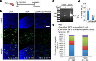
Targeted genome editing via engineered nucleases is an exciting area of biomedical research and holds potential for clinical applications. Despite rapid advances in the field, in vivo targeted transgene integration is still infeasible because current tools are inefficient1, especially for non-dividing cells, which compose most adult tissues. This poses a barrier for uncovering fundamental biological principles and developing treatments for a broad range of genetic disorders2. Based on clustered regularly interspaced short palindromic repeat/Cas9 (CRISPR/Cas9)34 technology, here we devise a homology-independent targeted integration (HITI) strategy, which allows for robust DNA knock-in in both dividing and non-dividing cells in vitro and, more importantly, in vivo (for example, in neurons of postnatal mammals). As a proof of concept of its therapeutic potential, we demonstrate the efficacy of HITI in improving visual function using a rat model of the retinal degeneration condition retinitis pigmentosa. The HITI method presented here establishes new avenues for basic research and targeted gene therapies.


Autores:

Suzuki, K., Tsunekawa, Y., Hernandez-Benitez, R., Wu, J., Zhu, J., Kim, E., Hatanaka, F., Yamamoto, M., Araoka, T., Li, Z., Kurita, M., Hishida, T., Li, M., Aizawa, E., Chen, S., Goebl, A., Soligalla, R.D., Qu, J., Jiang, T., Skowronska-Krawczyk, D., Rodriguez Esteban, C., Lajara, J., Nuñez, E., Guillen, P., Campistol, J.M., Matsuzaki, F., Liu, G.-H., Magistretti, P., Zhang, K., Callaway, E., Zhang, K and Izpisua Belmonte, J.C. “In vivo genome editing via CRISPR-Cas9 mediated homology-independent targeted integrationNature 540, 144-149 (01 December 2016).


Link: http://www.nature.com/nature/journal/v540/n7631/full/nature20565.html


----------------------

In vivo amelioration of age-associated hallmarks by partial reprogramming

Publicado en: CELL

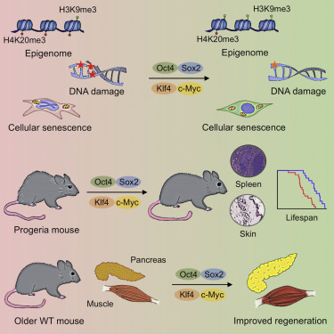
Aging is the major risk factor for many human diseases. In vitro studies have demonstrated that cellular reprogramming to pluripotency reverses cellular age, but alteration of the aging process through reprogramming has not been directly demonstrated in vivo. Here, we report that partial reprogramming by short-term cyclic expression of Oct4, Sox2, Klf4, and c-Myc (OSKM) ameliorates cellular and physiological hallmarks of aging and prolongs lifespan in a mouse model of premature aging. Similarly, expression of OSKM in vivo improves recovery from metabolic disease and muscle injury in older wild-type mice. The amelioration of age-associated phenotypes by epigenetic remodeling during cellular reprogramming highlights the role of epigenetic dysregulation as a driver of mammalian aging. Establishing in vivo platforms to modulate age-associated epigenetic marks may provide further insights into the biology of aging.


Referencia:

Alejandro Ocampo, Pradeep Reddy, Paloma Martínez-Redondo, Aida Platero-Luengo, Fumiyuki Hatanaka, Tomoaki Hishida, Mo Li, David Lam, Masakazu Kurita, Ergin Beyret, Toshikazu Araoka, Eric Vázquez-Ferrer, David Donoso, José Luis Román , Jinna Xu, Concepción Rodríguez-Esteban, , Gabriel Núñez, Estrella Nuñez Delicado,   Josep María Campístol, Isabel Guillén, Pedro Guillén and Juan Carlos Izpisua Belmonte. “In vivo amelioration of age-associated hallmarks by partial reprogramming” Cell 167, 1-15. December 15, 2016.


Link: http://www.cell.com/cell/abstract/S0092-8674(16)31664-6 _returnURL=http%3A%2F%2Flinkinghub.elsevier.com%2Fretrieve%2Fpii%2FS0092867416316646%3Fshowall%3Dtrue

----------------------

Intraarticular administration of Dobesilate for the Treatment of Knee Rheumatoid Arthritis

Publicado en: M.OJ Clin Med Case Rep


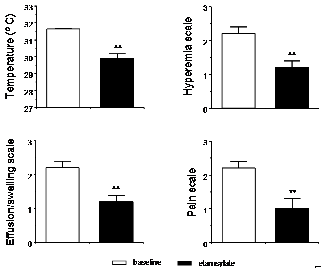
Rheumatoid arthritis is an inflammatory condition that causes a negative effect in patient well-being. Fibroblast growth factor participates in the pathology of rheumatoid arthritis through its proliferative, inflammatory and angiogenic activities. Dobesilate, an inhibitor of fibroblast growth factor shows marked anti-proliferative, anti-inflammatory, and anti-angiogenic activities. Our study shows that intraarticular application of dobesilate is effective in reducing symptoms and signs in patients with chronic knee rheumatoid arthritis.


Referencia:

Cuevas P, Fernández-Jaén T, Guillen P, Angulo J, Giménez-Gallego G (2016) “Intraarticular administration of Dobesilate for the Treatment of Knee Rheumatoid Arthritis”. M.OJ Clin Med Case Rep 5(1):00119 (2016)

Link: http://medcraveonline.com/MOJCR/MOJCR-05-00119.php

----------------------

3D Culture Supports Long-Term Expansion of Mouse and Human Nephrogenic Progenitors.

Publicado en: Cell Stem Cell

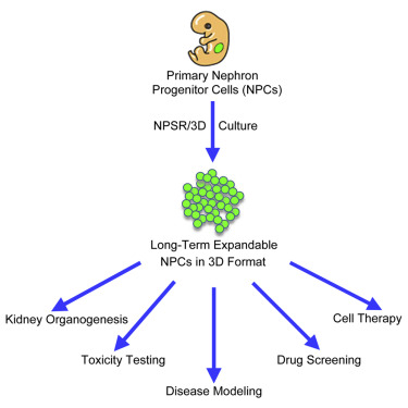
Transit-amplifying nephron progenitor cells (NPCs) generate all of the nephrons of the mammalian kidney during development. Their limited numbers, poor in vitro expansion, and difficult accessibility in humans have slowed basic and translational research into renal development and diseases. Here, we show that with appropriate 3D culture conditions, it is possible to support long-term expansion of primary mouse and human fetal NPCs as well as NPCs derived from human induced pluripotent stem cells (iPSCs). Expanded NPCs maintain genomic stability, molecular homogeneity, and nephrogenic potential in vitro, ex vivo, and in vivo. Cultured NPCs are amenable to gene targeting and can form nephron organoids that engraft in vivo, functionally couple to the host’s circulatory system, and produce urine-like metabolites via filtration. Together, these findings provide a technological platform for studying human nephrogenesis, modeling and diagnosing renal diseases, and drug discovery.


Referencia:

Zhogwei Li, Toshikazu Araoka, Jun Wu, Hsin-Kai Liao, Mo Li, Marta Lazo, Bing Zhou, Yinghui ui, Min-Zu Wu, Isao Tamura, Yu Xia, Ergin Beyret, Taiji Matsusaka, Ira Pastan, Concepcion Rodriguez Esteban, Isabel Guillén, Pedro Guillén, Josep M. Campistol, and Juan Carlos Izpisúa Belmonte. “3D Culture Supports Long-Term Expansion of Mouse and Human Nephrogenic Progenitors. Cell Stem Cell (2016), volume 19, Issue 4, p516, 6 October 2016


Link: http://www.cell.com/cell-stem-cell/abstract/S1934-5909(16)30211-9?_returnURL=http%3A%2F%2Flinkinghub.elsevier.com%2Fretrieve%2Fpii%2FS1934590916302119%3Fshowall%3Dtrue

----------------------

Identifying injury: “A retrospective cohort study based on information and communication technology”.

Publicado en: Archives of Orthopaedic and Trauma Surgery

Referencias:

Maryem I.; Bejarano, P; Villafañe, J.H, Ph.D, Msc; Lopez-Alcorocho, J.M.; Fernandez-Jaén, T.F., Guillén-Vicente, I.; Guillén-Garcia, P. “Identifying

injury: “A retrospective cohort study based on information and communication technology”. Archives of Orthopaedic and Trauma Surgery

----------------------

Case Report: Efficacy of dobesilate in insertional Achilles tendinopathy

Publicado en: F1000research

Leer más:

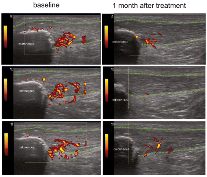
Achilles tendinopathy is an overuse syndrome, common among runners, with sometimes considerable negative impact on their performance, overall health, and well-being. Our report shows that local injection of an aqueous solution of the diethylammonium salt of dobesilate, an inhibitor of fibroblast growth factor with significant anti-angiogenic and anti-inflammatory effects, is effective in reducing vascular density and pain in insertional Achilles tendinopathy.


Referencia:

Cuevas P., Fernández-Jaen, T., Guillén, P. Angulo, J.y Giménez-Gallego, G. “Case Report:Efficacy of dobesilate in insertional Achilles tendinopathy” F1000research (2016), doi: 10.12688/f1000research.7260.1


Link: https://f1000research.com/articles/5-34/v1

----------------------

Viability of pathologic cartilage fragments as a source for autologous chondrocyte

Publicado en: Cartilage


OBJECTIVE:

To study if a culture of chondrocytes can be obtained from pathologic hyaline cartilage (PHC) fragments.

DESIGN:

Twenty-five men and 9 women with osteochondritis dissecans (OCD) in 11 cases, arthrosis in 13 patients, and trauma in the remaining 10 cases were included. The PHC fragments and a small sample of the next healthy cartilage were extracted by arthroscopy. According to the appearance, the PHC samples were divided into fixed (3 cases), flapped (6 patients), or loose bodies (25 cases), depending on the attachment degree of the cartilage to the subchondral bone. Approximately half of each pathologic sample and the whole healthy one were digested to isolate the cells trying to establish the cell culture.

RESULTS:

We were able to establish a cell culture in 7 out of 34 (20.6%) PHC samples (positive samples), whereas in the remaining 27 (79.4%) no cell growth was observed (negative samples). Most of the negative samples were loose bodies (P = 0.005) taken from patients with OCD or arthrosis (P = 0.001) with an evolution time of more than 1 year (P < 0.001). The best binary logistic regression model (P < 0.001) showed that the only factor affecting the establishment of cell culture was the evolution time (P = 0.044).

CONCLUSION:

It is possible to culture chondrocytes from osteochondral fragments if they are traumatic, within a year of injury and not from fragments due to arthrosis or OCD.


Referencia:

Guillen García, P., Rodriguez Iñigo, E. Guillén Vicente, I, Guillén Vicente, M., Fernández Jaén, T. Concejero López, V. Val, Maestro, A. Abelow, S. y López-Alcorocho, J.M. “Viability of pathologic cartilage fragments as a source for autologous chondrocyte” Cartilage  Vol. 7(2) 149-156 DOI: 10.1177/1947603515621998. SAGE (2016).


Link: https://www.ncbi.nlm.nih.gov/pmc/articles/PMC4797241/


----------------------

Nuestra experiencia con la técnica de implante de condrocitos autólogos para el tratamiento de lesiones condrales: resultados de 50 pacientes a 2 años de seguimiento

Publicado en: Rev. Esp. Artroscopia y Cirugía Articular.


Aim

The aim of this work was to evaluate the clinical results of the matrix-induced autologous chondrocyte implantation (MACI) technique on chondral defects of the knee and ankle. A study was conducted on 150 patients treated with MACI (performed by the same team of surgeons in all cases) during 2002-2007.

Methods

The lesion was evaluated by arthroscopy, and a cartilage biopsy was taken. The cellular implant was performed by arthroscopy in 53 patients, and by open surgery in the remaining cases. The results were measured following an in-house clinical protocol. Data corresponding to a mean follow-up period of 24 months were available in 50 out of the 150 implanted patients. Histological studies were performed in a second biopsy taken by arthroscopy in 5 patients 2 years after cell implantation.

Results

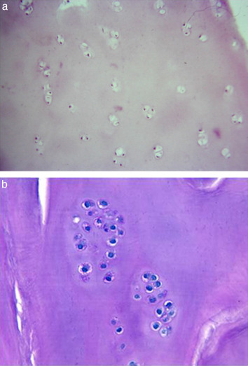
In 89% of patients without previous surgeries, and in 66% of patients with previous surgeries, the results were good or excellent for pain and motion, and most of them were able to return to their previous physical activities, including professional sport. Histological studies performed on a second biopsy showed that the new tissue is hyaline-like cartilage with lower cell numbers and subjectively softer than normal cartilage.

Conclusion

Arthroscopic MACI is a promising procedure to treat chondral lesions in the knee and ankle, although the histological analysis suggests that increasing cellular dosage would improve the results of the technique.

Clinical relevance

The importance of this study is that it presents an increased number of patients with chondral lesion treated with MACI. A high number of these patients were treated with arthroscopic MACI. In the present study it is shown that the treatment gives good results.


Referencia:

Guillen Garcia, P., Rodriguez Iñigo, E., Arauz de Robles, S., Guillén Vicente, M. Guillen Vicente, I. Caballero Santos, R., Ramos Martin, T., Fernández Jaén, T. Abelow, S. y J.M. López-Alcorocho. “Nuestra experiencia con la técnica de implante de condrocitos autólogos para el tratamiento de lesiones condrales: resultados de 50 pacientes a 2 años de seguimiento”. Rev. Esp. Artroscopia y Cirugía Articular.  22(3):120-125 (2015)


Link: http://www.sciencedirect.com/science/article/pii/S2386312915000833

----------------------