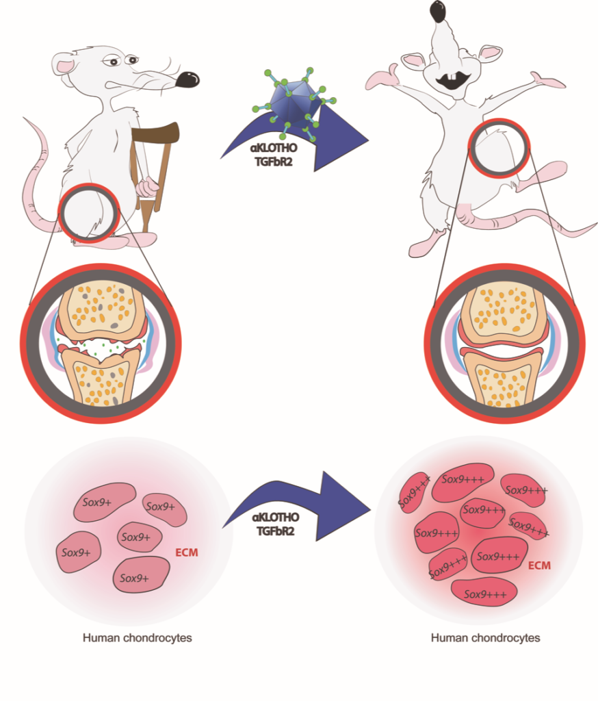
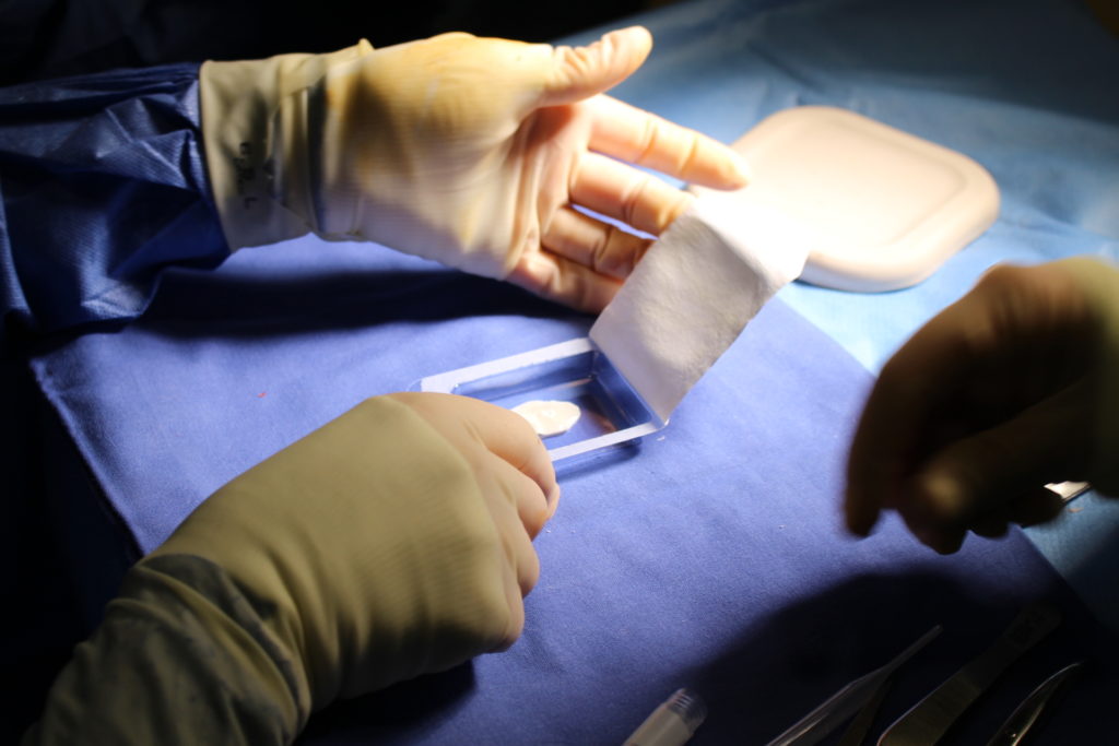
Fundacion

Fundacion

Evolución clínica y presencia de edema óseo subcondral a los dos años de tratamiento con implante de condrocitos autólogos de alta de densidad en la rodilla

Publicado en: Nature Communications

Abstract. Antecedentes y objetivo: Recientemente se ha descrito una nueva modalidad de la técnica
de implante de condrocitos autólogos sobre membrana de colágeno i/iii llamada HD-ACI (High
Density Autologous Chondrocyte Implantation) que está basada en el aumento de la densidad
celular. El objetivo de este trabajo fue estudiar la evolución clínica y la incidencia de la aparición
de edema óseo en pacientes con lesiones de cartílago en la rodilla tratados con HD-ACI al año
y a los 2 años de la intervención.
Métodos: Se trata de un estudio retrospectivo en 40 pacientes con lesiones condrales grado iii-iv.
Todos los pacientes fueron tratados con HD-ACI con una dosis celular de 5
× 106 condrocitos/cm2 de lesión. La percepción subjetiva de la mejora de los síntomas/funcionalidad se valoró mediante la escala del Comité Internacional de Documentación de la Rodilla (IKDC, International
Knee Documentation Committee). La presencia de edema óseo se evaluó a los 6, 12 y 24 meses de seguimiento por resonancia magnética. Comité Internacional de Documentación de la Rodilla (IKDC)
Resultados: Los valores de IKDC mostraron una mejoría significativa a los 12 y 24 meses
(p < 0,001). La diferencia media de IKDC entre la visita basal y los 12 meses fue de 26,3 puntos
y de 31,6 puntos a los 24 meses. El 27,5% de los pacientes presentaron edema óseo subcondral a los 2 años de seguimiento.
Conclusiones: HD-ACI es un tratamiento efectivo y seguro que mejora el dolor, la percepción
clínica y la funcionalidad de la articulación. No se ha encontrado correlación entre la presencia
de edema óseo y la evolución clínica de los pacientes.

Referencia:

López-Alcorocho JM, Aboli L, Rodríguez-Iñigo E, Guillén-Vicente I, Guillén-Vicente M, Caballero R, Casqueiro M, Fernández-Jaen TF, Abelow S, Guillen-García P. Clinical outcome and subchondral bone edema presence at two-year follow-up after high density autologous chondrocyte implantation treatment in the knee. Rev Esp Cir Ortop Traumatol. 2019, 63:253-260.

Link: https://doi.org/10.1016/j.recot.2019.03.004

----------------------

¿Constituye un cuerpo libre articular una buena fuente de células para un implante de condrocitos autólogos?

Publicado en: Nature Communications

Abstract. El empleo de cuerpos libres articulares como fuente de células para el cultivo de un futuro
implante de condrocitos sigue siendo controvertido. El objetivo del trabajo fue estudiar
si podían obtenerse condrocitos viables para cultivo in vitro de fragmentos desprendidos
de cartílago de 50 pacientes afectados de osteocondritis disecante, artrosis
o traumatismo. Las células se aislaron de los fragmentos desprendidos para estudiar
su capacidad de establecer un cultivo in vitro. También se realizó análisis histológico
y molecular de las muestras objeto de estudio. Pudimos establecer un cultivo celular
solo en el 22% de los fragmentos desprendidos analizados, mientras que en el 78%
no se observó crecimiento celular. La mayoría de las muestras en las que no se logró el
cultivo celular eran cuerpos libres tomados de pacientes con osteocondritis disecante
o artrosis (p = 0,002) con un tiempo de evolución mayor de 1 año (p <0,001). Los
resultados del análisis histológico y molecular confirmaron la presencia de cartílago
hialino solo en las muestras en las que pudo establecerse el cultivo. Nuestro estudio
demuestra que no en todos los casos se consigue el cultivo celular, y que el tiempo de
evolución es el factor principal que influye en ello.

Referencia:

López-Alcorocho JM, Rodríguez-Iñigo E, Guillén-Vicente I, Caballero-Santos R, Guillén-Vicente M, Casqueiro-Abad M, Fernández-Jaén TF, Concejero V, Abelow S, Guillén-García P. ¿Constituye un cuerpo libre articular una buena fuente de células para un implante de condrocitos autólogos? Journal of Cartilage Disease 2019.

----------------------

Stabilization of heterochromatin by CLOCK promotes stem cell rejuvenation and cartilage regeneration.

Publicado en: Nature Communications

Abstract. Accumulating evidence indicates an association between the circadian clock and the aging process. However, it remains elusive whether the deregulation of circadian clock proteins underlies stem cell aging and whether they are targetable for the alleviation of aging-associated syndromes. Here, we identified a transcription factor-independent role of CLOCK, a core component of the molecular circadian clock machinery, in counteracting human mesenchymal stem cell (hMSC) decay. CLOCK expression was
decreased during hMSC aging. In addition, CLOCK deficiency accelerated hMSC senescence, whereas the overexpression of CLOCK, even as a transcriptionally inactive form, rejuvenated physiologically and pathologically aged hMSCs. Mechanistic studies revealed that CLOCK formed complexes with nuclear lamina proteins and KAP1, thus maintaining heterochromatin architecture and stabilizing repetitive genomic sequences. Finally, gene therapy with lentiviral vectors encoding CLOCK promoted cartilage
regeneration and attenuated age-related articular degeneration in mice. These findings demonstrate a noncanonical role of CLOCK in stabilizing heterochromatin, promoting tissue regeneration, and mitigating aging-associated chronic diseases.

Referencia:

Liang C, Liu Z, Song M, Li W, Wu Z, Wang Z, Wang Q, Wang Si, Yan K, Sun L, Hishida T, Cai Y, Izpisua Belmonte JC, Guillen P, Chan P, Zhou Q, Zhang W, Qu J, Liu GH. Stabilization of heterochromatin by CLOCK promotes stem cell rejuvenation and cartilage regeneration. Cell Res 2020 Jul;31(2):187-205. doi: 10.1038/s41422-020-0385-7

Link: https://doi.org/10.1038/s41422-020-0385-7

----------------------

Histology and Type I Collagen, Tenascin and Elastin Expression in Autologous and Allogeneic Anterior Cruciate Ligamentoplasties.

Publicado en: Nature Communications

Abstract. Background. Ligamentization has been accepted in Anterior Cruciate Ligament (ACL)
surgery. The purpose of this study is to evaluate ligamentization of different allografts
and autografts used as plasties through histological and type I collagen (COL1), tenascin
(TEN) and elastin (ELA) expression analysis.
Methods. Prospective study of patients who underwent ACL reconstruction with at least
1-year follow-up. Biopsies were taken by arthroscopy and tissue sections were stained with
hematoxilin-eosin and Masson’s trichrome. Relative COL1, TEN and ELA expression was
studied by RT-PCR. Hamstring tendon and intact ACL biopsies were used as controls.
Results. Eleven patients with a mean age of 36.6 ± 12.0 years were included. In 9 cases,
ACL reconstruction was carried-out with autologous hamstring tendons while in the 2
remaining patients ACL was reconstructed using an achilles allograft. COL1, TEN and
ELA expression of plasties was significantly different to ACL but similar to hamstring
tendon. Histological analysis showed a dense fibroconnective tissue with cells similar to
tenocytes, which could not be classified neither as a tendon nor as a ligament.
Conclusions. An ACL plasty, independently of its origin (autologous hamstring or allogeneic
achilles) does not become a ligament, at least in our study period (5 years) but it does
maintain ACL functionality.

Referencia:

Guillén-Vicente I, López-Alcorocho JM, Salvador-Marín J, Rodríguez-Iñigo E, Díaz-Motola G, Fernández-Jaén TF, Ibáñez E, Guillén-García P. Histology and type I collagen, tenascin and elastin expression in autologous and allogeneic anterior cruciate ligamentoplasties. Muscle, Ligaments and Tendons Journal 2020 10 (3):473-480.

----------------------

Traumatología INVESTIGACIÓN Reparación anatómica de las lesiones del tendón distal del bíceps braquial utilizando la técnica quirúrgica de doble incisión.

Publicado en: Nature Communications

Abstract. En este trabajo se estudió la evolución clínica de pacientes con roturade tendón del bíceps braquial tratados mediante reconstrucción quirúrgica según nuestra propiatécnica de doble incisión.Pacientes: Se incluyó a 20 pacientes (21 casos) de rotura de tendón del bíceps braquial trata-dos mediante nuestra propia técnica quirúrgica de doble abordaje modificada, consistente en2 incisiones mínimas. La evaluación funcional se determinó mediante la escala Mayo Elbow Per-formance Score, la fuerza muscular se determinó con el cuestionario Medical Research Councily el dolor mediante la Escala Analógica Visual a los 12 meses de seguimiento. Se determinó elrango de movilidad midiendo los grados de flexión y extensión.Resultados: Un a˜no después de la cirugía todos los pacientes habían vuelto al deporte al mismonivel que tenían previamente. La puntuación de Escala Analógica Visual fue de 1,90 ± 0,89(media ± desviación estándar) y la mediana de la escala Mayo Elbow Performance Score de 90(mínimo-máximo: 80-100). Del total, 16 (76,2%) presentaron la máxima puntuación en la escalaMedical Research Council (puntuación 5), mientras que 5 casos (23,81%) tuvieron una puntaciónde 4+. La media de extensión fue de 136,67◦(IC 95%: 134,14◦-139,19◦) y −7,38◦de flexión(IC 95%: [−10,04◦]-[−4,72◦]).

Referencia:

Ibánez E, Araiza E, Rodríguez-Iñigo E, Guillén-García P, López-Alcorocho JM. Reparación anatómica de las lesiones del tendón distal del bíceps braquial utilizando la técnica quirúrgica de doble incisión. Revista Española de Cirugía Ortopédica y Traumatología 2021 Feb 25;S1888-4415(21)00005-9. doi: 10.1016/j.recot.2020.10.005.

Link: https://doi.org/10.1016/j.recot.2020.10.005

----------------------

High-Density Autologous Chondrocyte Implantation (HD-ACI) in patients with bilateral knee chondral defects

Publicado en: Nature Communications

Abstract. Bilateral knee chondral defects can be treated in one or two surgical acts.
Objectives: To study clinical outcome of patients with bilateral knee chondral defects treated with
High-Density Autologous Chondrocyte Implantation (HD-ACI) during the same surgery.
Methods: Study on eight patients (4 women and 4 men) with chondral defects in both knees
treated with HD-ACI during the same surgical act. Patients were evaluated 2, 6, 12 and 24 months
post-op for treatment safety and efficacy: visual analogic scale (VAS) for pain, International Knee
Documentation Committee (IKDC) for subjective patient’s perception of both knees and EuroQol
five-dimensional five-level questionnaire (EQ-5D-5 L) for quality of life. Integrity of neoformed
tissue at 12 and 24-month follow-up was assessed by the magnetic resonance observation of
cartilage repair tissue (MOCART) score.
Results: VAS decreased from baseline to 24 months from a median of 8 (6 – 9) to a median of 0 (0
– 5) ( P < .001). IKDC increased from a median value of 39.0 (17.2 – 48.3) in the basal visit to a
median of 83.7 (24.0 – 98.0) in the 24-month visit ( P < .001). EQ-5D-5 L decreased at 2 months
post-op and increased throughout the follow-up visits thereafter, needing 24 months after surgery
to reach a value higher than the basal one. Median MOCART at 12 months was 80.3 (58.0 – 89.7)
and 81.3 (54.0 – 89.9) at 24 months.

Referencia:

Guillén-Vicente I, López-Alcorocho JM, Rodríguez-Iñigo E, Guillén-Vicente M, Fernández-Jaén TF, Cortés JM, Abelow S, Guillén-García P. High-Density Autologous Chondrocyte Implantation (HD-ACI) in patients with bilateral knee chondral defects. Journal of Cartilage & Joint Preservation TM 1 (2021) 100013

Link: https://doi.org/10.1016/j.jcjp.2021.100013

----------------------

In vivo partial cellular reprogramming enhances liver plasticity and regeneration.

Publicado en: Nature Communications

Abstract. Mammals have limited regenerative capacity, whereas some vertebrates, like fish and salamanders, are able to regenerate their organs efficiently. The regeneration in these species depends on cell dedifferentiation followed by proliferation. We generate a mouse model that enables the inducible
expression of the four Yamanaka factors (Oct-3/4, Sox2, Klf4, and c-Myc, or 4F) specifically in
hepatocytes. Transient in vivo 4F expression induces partial reprogramming of adult hepatocytes
to a progenitor state and concomitantly increases cell proliferation. This is indicated by reduced
expression of differentiated hepatic-lineage markers, an increase in markers of proliferation and
chromatin modifiers, global changes in DNA accessibility, and an acquisition of liver stem and
progenitor cell markers. Functionally, short-term expression of 4F enhances liver regenerative
capacity through topoisomerase2-mediated partial reprogramming. Our results reveal that liverspecific 4F expression in vivo induces cellular plasticity and counteracts liver failure, suggesting
that partial reprogramming may represent an avenue for enhancing tissue regeneration.

Referencia:

Hishida T, Yamamoto M, Hishida-Nozaki Y, Shao C, Huang L, Wang C, Shojima K, Xue Y, Hang Y, Shokhirev M, Memczak S, Kumar Sahu S, Hatanaka F, Rabadan Ros R, Maxwell MB, Chavez J, Shao Y, Liao HK, Martinez-Redondo P, Guillen-Guillen I, Hernandez-Benitez R, Rodriguez Esteban C, Qu J, Holmes MC, Yi F, Hickey RD, Guillen Garcia P, Nuñez Delicado E, Castells A, Campistol JM, Yu Y, Hargreaves DC, Asai A, Reddy P, Liu GH, Izpisua Belmonte JC. In vivo partial cellular reprogramming enhances liver plasticity and regeneration. Cell Rep. 2022 Apr 26;39(4):110730. doi: 10.1016/j.celrep.2022.110730.

Link: http://creativecommons.org/licenses/by/4.0/

----------------------

High density-autologous chondrocyte implantation for the treatment of bilateral ankle cartilage defects: Report of two cases.

Publicado en: Nature Communications

Abstract. To study clinical outcome of patients with bilateral ankle chondral defects treated with High-Density
Autologous Chondrocyte Implantation (HD-ACI) in the same surgical act.
Cases presentation: Two men (27 and 48 years-old) with chondral defects in both ankles evidenced by MRI. Chondral
lesions were treated with HD-ACI in the same surgical act and anesthesia, consecutively. Patients were evaluated
2, 6 and 12 months after surgery to check for treatment safety and efficacy. Pain, evaluated by the Visual
Analogic Scale (VAS), decreased from very high values (8.5 – 9) to normal or almost scores one year after surgery (0-1). Ankle functionality measured by the American Orthopedic Foot & Ankle Society score (AOFAS) and quality of life evaluated by the EuroQol five-dimensional five-level questiionnaire (EQ-5D-5L) behaved similarly to pain. Thus, both parameters increased and/or slightly decreased at 2 months (AOFAS socre: 90 and 89; EQ-5D-5L score: 0.89 and 0.91). MRI from both ankles revelaed that chondral defects were filled with a material of similar aspect to the surrounding cartilage.

Referencia:

Guillen-Vicente I, Lopez-Alcorocho JM, Rodríguez-Iñigo E, Navarro R, Guillén-Vicente M, Fernández-Jaen T, Guillén-García P. High density autologous chondrocyte implantation for the treatment of bilateral ankle cartilage defects: Report of two cases. Foot and ankle surgery: Techniques reports and cases 2 (2022) 100218

Link: https://doi.org/10.1016/j.fastrc.2022.100218

----------------------

The use of high-density autologous chondrocytes implant for the treatment of hip joint chondral defects.

Publicado en: Nature Communications

Abstract. In this work we describe the use of high-density autologous chondrocyte implantation
(HD-ACI) to treat 2 patients with chondral defects in the hip.
Case Presentation: Two male, 35- and 37-year-old, with chondral lesions and femoroacetabular
impingement (FAI) were included. Chondrocytes were isolated expanded in vitro and then implanted
at a density of 5 million cells per cm 2 onto a type I/III porcine collagen membrane. None
of the patients had surgery-derived complications. Pain disappeared in both cases at 6 months
after surgery and was maintained until follow-up end (36 months). International Hip Outcome
Tool (iHOT) score increased at 6 and 12 months and then leveled off until month 36.Three-year
arthro-MRI showed the cartilage defects had been filled with a material similar to the surrounding
healthy cartilage.
Conclusion: HD-ACI is a safe and effective technique for focal cartilage lesion treatment in patients
affected by evolved FAI.

Referencia:

Torres-Eguia R, Lopez-Alcorocho JM, Rodriguez-Iñigo E, Guillen-Vicente I, Guillen-Vicente M, Diaz-Navarro ME, Aboli L, Fernández-Jaén TF, Guillen-Garcia P. The use of high-density autologous chondrocytes implant for the treatment of hip joint chondral defects. Journal of Cartilage & Joint preservation 2022

Link: https://doi.org/10.1016/j.jcjp.2022.100070

----------------------

FUNDACIÓN DR. PEDRO GUILLÉN Y LÍNEA DIRECTA LLEGAN A UN ACUERDO PARA IMPULSAR LA INVESTIGACIÓN EN EL CAMPO DE LA TRAUMATOLOGÍA

Dentro de su apuesta por la investigación en el ámbito de las Ciencias de la Salud, la Fundación Doctor Pedro Guillén ha llegado a un acuerdo con Línea Directa Aseguradora para apoyar la investigación “Nuevo concepto de mapas multiómicos del músculo esquelético humano”.

Este importante proyecto profundizará en la obtención de una visión global del músculo esquelético humano con el objetivo de brindar al paciente una rehabilitación rápida y efectiva, mejorando su calidad de vida y una mayor autonomía personal. Para ello, la investigación se centrará en la comprensión de los cambios dinámicos del músculo esquelético, proporcionando una visión completa de la función muscular en el ser humano.

 [···]

----------------------